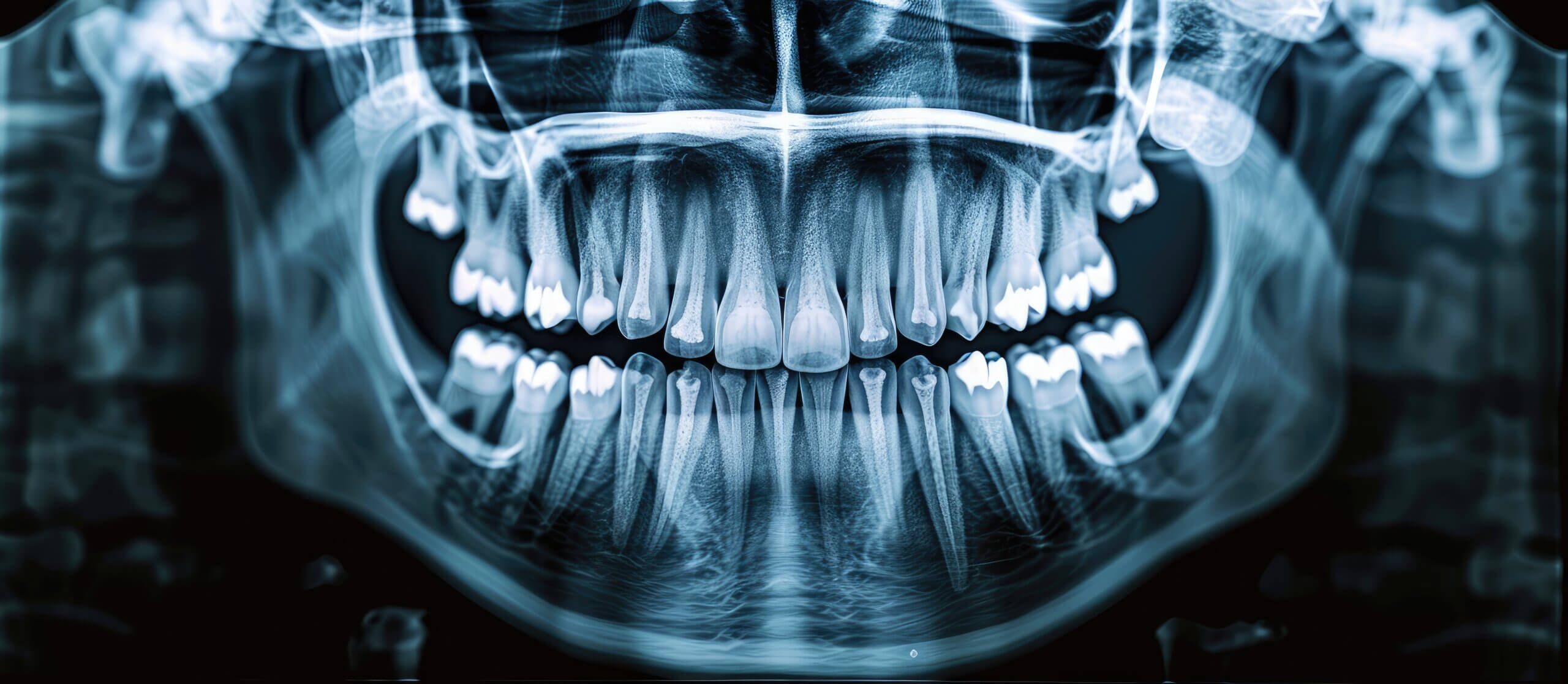
The X-Ray Report: Understanding Your Dental Imaging
Dental X-rays can be valuable for diagnosing and treating various oral health conditions. Despite common misconceptions, routine X-rays involve few risks, which makes obtaining images of your teeth and jaws safe. Before your next checkup, learn about dental imaging, what these images show, and the dental X-rays used to evaluate oral health.
How are Dental X-rays taken?
When you visit your Melbourne, FL, dentist for a routine checkup or treatment, she may suggest dental X-rays to examine your teeth and oral structures better. First, the technician will cover you with a lead apron to protect your body from radiation. A thyroid collar may also be wrapped around your neck.
Next, you’ll be positioned in a dental chair or asked to stand before the X-ray machine. A small plastic apparatus is inserted into your mouth, and you’ll be asked to bite down gently to hold it in place. The technician will then position the X-ray machine, and an image of your mouth will be taken with a button click. Alternatively, the machine may rotate around your head for approximately 15 seconds.
What Do Dental X-Rays Show?
A dental X-ray can show many dental issues with the mouth’s teeth, gums, and bones. From cavities between teeth to the position of the wisdom teeth, dental X-rays are an essential diagnostic tool dentists use to help them make informed treatment recommendations.
A dental X-ray can also show bone loss, tooth infections, impacted teeth, occlusion problems, cysts, tumors, and problems with restorations like crowns, fillings, and implants.
What are the Different Types of Dental X-Rays?
Similar to radiographs taken of other body parts, dental X-rays use electromagnetic radiation to obtain detailed pictures of your mouth. The two primary types of dental X-rays include intraoral and extraoral X-rays.
Intraoral X-Rays
Intraoral dental X-rays refer to X-rays that are taken when the sensor or film is inserted inside the mouth. The most common types of intraoral X-rays include:
- Bitewing – X-rays show the upper and lower teeth in a designated mouth area. This radiograph can help show decay between the teeth or below the gumline.
- Periapical—A periapical X-ray shows the entirety of a tooth, from the root tip to the crown. It can detect tooth decay, gum disease, bone loss, and other abnormalities.
- Occlusal – Occlusal dental X-rays are generally used to detect problems on the roof or floor of the mouth. This is especially beneficial for identifying impacted or fractured teeth.
Extraoral X-Rays
Extraoral dental X-rays are radiographs taken with the sensor or film outside the mouth. Some of the most common examples of extraoral X-rays include:
- Panoramic – Panoramic X-rays show all structures inside the mouth in a single image, including the upper and lower arch, nerves, jaw joints, sinuses, and all supporting bones.
- Cephalometric – A cephalometric X-ray captures the entire head from one side, allowing your dentist to see the location of your teeth about your jaw. Orthodontists most commonly use this type of dental X-ray during treatment.
- Cone Beam CT (CBCT) – Cone beam CT X-rays, or CBCT, use computed tomography (CT) scans to obtain 3D images of the teeth, joints, jaws, sinuses, and nerves. This type of X-ray can also identify facial fractures and oral tumors.
Schedule Your Next Appointment
Dentists commonly take dental X-rays during new patient exams and routine checkups to identify potential oral health problems, such as tooth decay, growths, and bone loss. By revealing more about the health of your teeth through dental X-rays, your Melbourne, Florida dentist can make informed treatment decisions to help keep your smile healthy and beautiful. To schedule your next appointment, contact Artistic Touch Dentistry today at 321.724.1400.

